Insert Experiment — Abstract
Abstract submitted to ATC-ATG 2026 presenting the 3D-printed MPI mouse-bed insert as a multimodal registration solution, evaluating how well it improves MPI-to-CT registration accuracy over taped fiducials.
Presenting author, with Tyler Tesdall, Daniela Paola Valdés, and Dr. Carlos Rinaldi-Ramos — Rinaldi-Ramos Laboratory, University of Florida
Overview
Design of a 3D Printed Magnetic Particle Imaging Bed Insert for Multimodal Registration — an abstract submitted to ATC-ATG 2026 with Tyler Tesdall, Daniela Paola Valdés, and Dr. Carlos Rinaldi-Ramos, presenting the 3D-printed MPI mouse-bed insert from the companion Insert project as a multimodal registration solution and evaluating how well it performs against the taped-fiducial method it replaced.
Problem
Magnetic particle imaging (MPI) generates background-free signal from iron-oxide nanoparticles but provides no anatomical context on its own, so it must be registered to an anatomical modality like CT. Conventional workflows either image fiducials alongside the subject, where spillover can affect in vivo quantification (Shakeri-Zadeh et al., 2025), or acquire a separate fiducial-only scan. Fiducial preparation and placement are usually manual, taped fiducials shift between studies, and taping confines them to the bed surface rather than distributing them through the field of view, which is what minimizes registration error.
Goal
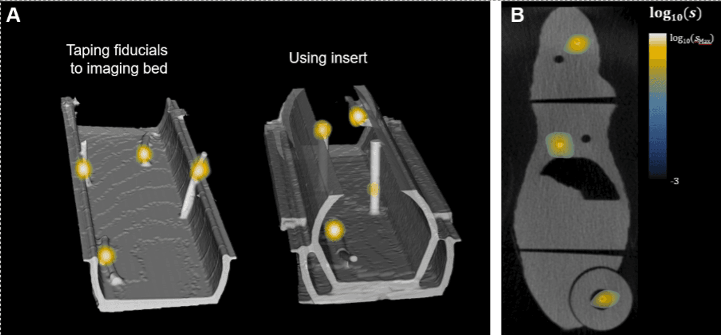
Evaluate whether a 3D-printed insert that reproducibly positions fiducials at CAD-defined 3D coordinates spanning the field of view, including positions elevated above the bed floor, improves MPI-to-CT registration agreement over taping fiducials directly to the bed.
Design Process
- Designed a clear resin insert in CAD to seat reproducibly in the lab's existing MPI mouse bed
- Positioned four capillary fiducial channels at defined 3D coordinates spanning the field of view, including elevated positions above the bed floor
- Filled fiducials with 2 µL of VivoTrax diluted 1:1 (v/v) in Omnipaque so they were visible in both MPI and CT
- Registered an anatomically correct 3D-printed mouse phantom (Valdés et al., 2026) in MPI and CT using both taped fiducials and the insert, then compared segmentation agreement between the two methods
Results
Compared agreement between MPI and CT mouse-phantom segmentations registered via taping versus the insert, to quantify whether distributing fiducials through the field of view improves registration accuracy over confining them to the bed surface.
What I Learned
Writing this abstract pushed me to quantify something I'd mostly reasoned about qualitatively while designing the insert: whether spreading fiducials through the field of view actually improves registration accuracy over taping them to the bed, not just whether it's more convenient to use.
Tools and Skills
Full Abstract
View or download the full abstract as a PDF.
View / Download Full Abstract